Imaging Tests for Neuroendocrine Tumors
Imaging tests are used to diagnose and assess neuroendocrine tumors. It can sometimes be difficult to image NETs. Your doctor may use different scans than those used for other cancer types.
 Computed Tomography (CT)
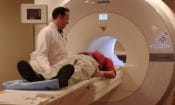
CT is an imaging technique that uses a highly specialized X-Ray machine and computers to create multiple cross-sectional images of the body. CT can generate images of different body tissues as well as help detect tumors. A dye may be injected into a vein or swallowed to help the organs or tissues show up more clearly.
Magnetic Resonance Imaging (MRI)
MRI uses radio waves, a powerful magnetic field, and a computer to generate detailed (2 or 3 dimensional) images of the body. These images are very useful in contrasting different types of tissue as well as detecting abnormal growths such as tumors within the body.
Somatostatin Receptor Scintigraphy (SRS)
Somatostatin Receptor Scintigraphy (SRS) is a type of radionuclide scan that uses a radioactive substance that can bind to a tumor’s somatostatin receptors and illuminate them. Not all tumors have somatostatin receptors.
Patients who take somatostatin analogs should discuss with their doctor the best timing for their exam as treatment can interfere with the test. Talk to your doctor about how to adjust your somatostatin analog before testing.
SRS can also help predict response to somatostatin analogs therapy as well as peptide receptor radionuclide therapy (PRRT).
Octreotide scan
A very small amount of radioactive octreotide is injected into a vein and travels through the bloodstream. Octreotide attaches to the tumor, and a special camera that detects radioactivity shows where the tumors are in the body.
Positron Emission Tomography (PET)
Positron Emission Tomography (PET) is another type of scan that uses radioactive material and a special scanning device to detect neuroendocrine tumors.
Ga-68 Dotatate PET

PET with Ga-68 dotatate is a newer SRS test that uses a radioactive tracer called Ga-68 dotatate to bind to a tumor’s somatostatin receptors. Following the injection of Ga-68 dotatate, patients undergo a PET scan. The images clearly show NETs anywhere in the body.
Copper Cu 64 Dotatate PET Scan
In 2020, the U.S. Food and Drug Administration approved a copper Cu 64 dotatate injection for use in positron emission tomography (PET) scans for certain kinds of neuroendocrine tumors (NETs). Diagnostic tracers like Cu 64 dotatate are used with PET scans and other functional imaging scans to improve how NETs are detected and staged.
Available under the commercial name DetectnetTM, the copper Cu 64 dotatate agent is a radiopharmaceutical tracer agent used in the localization of somatostatin receptor positive NETs in adults. Developed by RadioMedix Inc. and its commercial partner Curium, Detectnet is the first commercially available Cu 64 diagnostic agent on the U.S market.
According to phase 3 trials, Detectnet demonstrated clinical sensitivity to allow clinicians to develop more precise treatment approaches for their patients who have NETs. In addition, less than 2% of study participants in clinical trials experienced adverse reactions to the agent, which typically included nausea, vomiting, and flushing.
After FDA approval, the CU 64 dotatate injection was immediately made available through nuclear pharmacies. It’s 12.7-hour half-life allows Detectnet to be shipped throughout the US.
FDG-PET scan (fluorodeoxyglucose-positron emission tomography scan)
FDG-PET can find fast-growing neuroendocrine cancer cells for aggressive tumors. A small amount of FDG, a type of radioactive glucose (sugar), is injected into a vein. The PET scanner rotates around the body and makes a picture of where the body is using glucose. Cancer cells show up brighter in the picture because they are more active and take up more glucose than normal cells do. FDG is most commonly used to detect tumors that are high grade and/or poorly differentiated.
MIBG scan
An MIBG scan is used to find certain neuroendocrine tumors, such as pheochromocytoma and paraganglioma. A very small amount of a radioactive substance called MIBG is injected into a vein and travels through the bloodstream. The scan detects neuroendocrine tumor cells that take up MIBG. Scans may be taken over 1-3 days.
Endoscopy
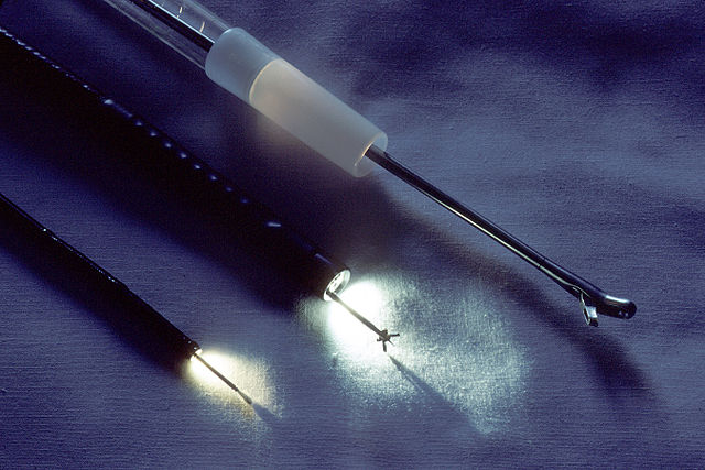 Endoscopy is a medical procedure that uses an endoscope to view the lining of multiple organs and tracts of the body. An endoscope is a flexible or rigid tube that has imaging capabilities and can enable small surgical procedures. Endoscopy can be used to visualize tumors in the lungs and gastrointestinal tract (stomach, small and large intestine and rectum). Sometimes an ultrasound probe is put on the end of the endoscope to perform what is called an endoscopic ultrasound (EUS), which can be particularly useful for looking at gut-adjacent organs like the pancreas.
Bronchoscopy
Bronchoscopy is a procedure to look inside the trachea and large airways in the lung for abnormal areas. A bronchoscope is a thin, tube-like instrument with a light and a lens for viewing. It may also have a tool to remove tissue samples to be checked under a microscope for signs of cancer. A physician inserts a bronchoscope through the nose or mouth into the trachea and lungs.

Tom Hope, MD, University of California, San Francisco
The basics of imaging for NETs Watch Video
Sumeet Virmani, MD, Rush University.
Ga-68 scan is gamechanger. Watch Video
Evelyne Loyer, MD, MD Anderson Cancer Center
What imaging test is used when. Watch Video