Interventional Radiology as a Treatment for Neuroendocrine Tumors
Hepatic embolization
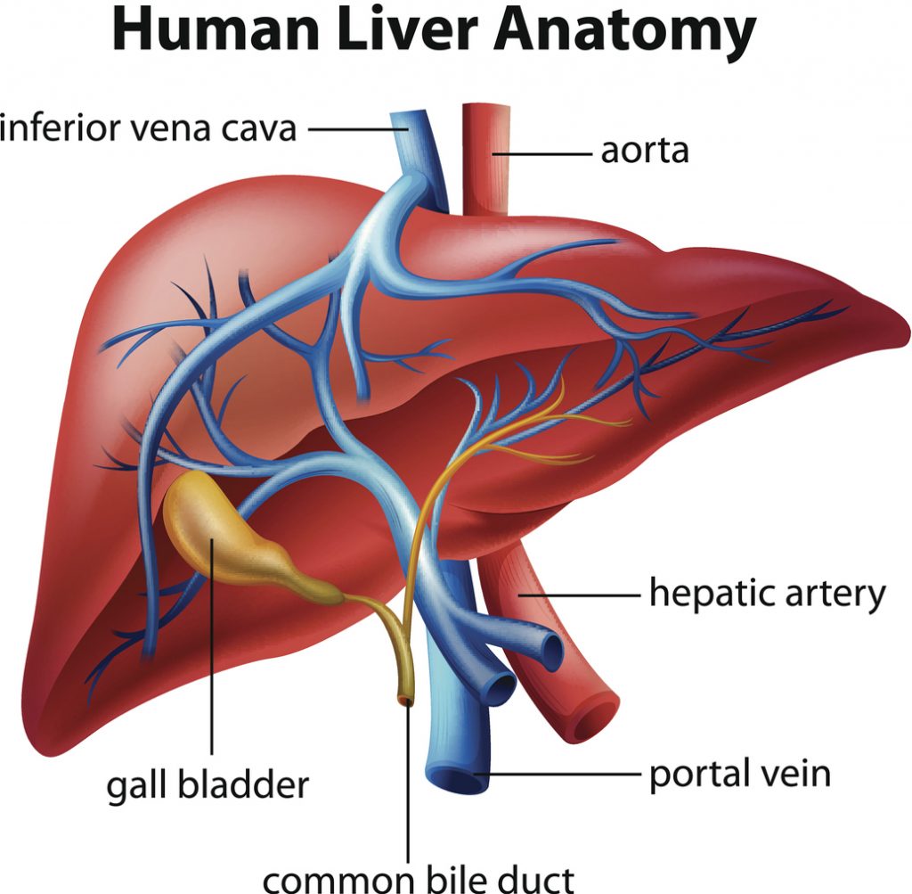
A hepatic embolization blocks or reduces flow of blood in the hepatic vessel to kill cancer cells growing in the liver. The portal vein continues to supply blood to the liver. There are two types of hepatic embolization:

- Bland embolization: the injection of embolic particles, such as tiny gelatin sponges or beads (to reduce blood flow).
- Chemoembolization (TACE): the injection of embolic particles (to reduce blood flow) mixed with chemotherapy (to kill tumor cells) or direct injection of chemotherapy into the artery using a catheter.
Radioembolization combines embolization with radiation therapy to kill tumor cells in the liver. The doctor injects millions of radioactive microspheres (microscopic beads) into the hepatic artery, which emit radiation to kill tumor cells.
Radiofrequency Ablation (RFA)
Radiofrequency ablation (RFA) uses electrical current to destroy tumor cells. RFA involves placing a small probe into a tumor. This raises the temperature of the tumor tissue and destroys it. RFA can be done laparoscopically but is more commonly done in combination with liver resection.

Riad Salem, MD, Northwestern University
The latest in liver-directed therapies. Watch Video
Karen Grace, Northwestern University.
What is it like to undergo interventional radiology treatment? Watch Video
David Agarwal, MD, Indiana University.
Understanding interventional radiology Watch Video